1. PERCUSSION IN RESPIRATORY SYSTEM :
PURPOSE :
(1) diagnostic – to determine the state of the underlying tissues
(2) topographical – to delineate the borders
METHOD :
Preferred position – sitting up.
Anterior chest wall – patient sits on a stool, with body bolt upright, completely relaxed, and with the
sides symmetrical.
Posterior chest wall – patient bend slightly forward with head flexed on the chest, the shoulders sagging,
and the arms resting, either crossed or uncrossed, on the thighs. During percussion of the interscapular
and scapular regions, the patient is directed to place his hands over the shoulders, after crossing the
arms in front of the chest.
Axillae – patient is instructed to put his hands over the head.
CARDINAL RULES :
1. The pleximeter, usually the middle finger of the examiner’s left hand must be firmly applied to the chest
wall, so that no air pockets are interposed in between.
2. The plessor, usually the middle finger of the examiner’s right hand, is kept flexed at a right angle and must hit the middle phalanx of
the pleximeter finger, perpendicular, with the pad and not the tip of the finger.
3. The percussion stroke must be sudden, the plessor finger being withdrawn immediately after the
stroke, to prevent a damping of the note. The movement of the percussion must originate at the wrist,
which is kept completely relaxed.
4. The force of the stroke must be varied according to the purpose of the percussion, the tissue or organ
being percussed, the thickness of the chest wall, the area of the chest wall percussed, the age, sex and
state of nutrition of the patient.
5. Percussion should proceed from resonant to dull areas or “more resonant” to “less resonant” areas.
6. When delineating the border of an organ, such as heart or liver, the long axis of the pleximeter finger must
be kept parallel to the expected position of that border.
7. It is better, whenever possible, to keep the pleximeter finger along an interspace.
8. The area percussed must be more or less equidistant from the two ears.
SPECIAL TECHNIQUES :
Flicking percussion – Special form of light percussion, the surface to be percussed being flicked with the
finger and the thumb. Useful in topographical percussion of the cardiac borders and for eliciting metallic
resonance in pneumothorax.
Palpatory percussion – Direct percussion with pads of the three middle fingers of the right hand over
posterior chest. Useful in detecting fluid or consolidation.
TYPE OF PERCUSSION NOTE CAUSATIVE LESION
Resonant Normal aerated lung
Tympanitic Superficial lung cavity, pneumothorax
Subtympany (boxy note) Above pleural effusion or consolidation
Hyperresonant Pneumothorax
Impaired Pulmonary fibrosis, sometimes consolidation or collapse
Dull Pulmonary consolidation or collapse, thickened pleura, tumour or raised diaphragm
Flat Pleural effusion
Stony dull Massive growth in the lung or pleura
Cracked-pot Lung cavity communicating with a bronchus
Amphoric Pneumothorax or large lung cavity
Bell tympany Massive pneumothorax
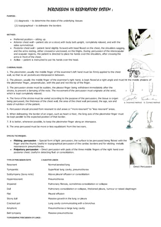
TOPOGRAPHIC PERCUSSION OF LUNGS :
Direct Percussion
a. Incorrect method b. Correct method
2. Apical percussion – Diminution or absence of the supraclavicular zone of resonance – pulmonary tuberculosis.
Increased extent of resonance, bilaterally – emphysema. Alternate method is mapping Kronig’s isthmus.
Basal percussion – Lower border of lung resonance is depressed - emphysema or pneumothorax, and
raised – lung fibrosis, collapsed lung, consolidation, ascites, massive abdominal tumour or pleural effusion.
Percussion of the mediastinum – To detect widening dullness on either side of sternum.
Tidal percussion – Percussion of lower border of lung resonance, on each side, at the height of deep inspiration and expiration –
to determine the extent of diaphragmatic excursion, pulmonary fibrosis.
SPECIAL PERCUSSIONAL FINDINGS IN DISEASE :
Reduction of both cardiac and liver dullness – hyperinflation of the lungs.
Shifting dullness – hydro and pyopneumothorax.
S- shaped curve of Ellis – moderate sized effusions within the pleural sac.
Grocco’s triangle (Paravertebral triangle of dullness) – Bounded
Medially - the mid-spinal line from the level of the effusion to the level of the tenth dorsal vertebra,
Below - a horizontal line extending outwards from the tenth dorsal vertebra along the lower limit of lung resonance, and
Laterally - a curved line connecting these two lines.
Seen in large or medium sized pleural effusion, over the back of the chest, on the opposite side of effusion.
Garland’s triangle – Lower relaxed part of lung in moderate or large pleural effusion is tympanitic or subtympanic.
Obliteration of Traube’s space – In left sided pleural effusion.
William’s tracheal resonance – Area of tympany over the first or second interspace, close to sternum.
Seen in patch of consolidation or fibrosis interposed between the trachea or a major bronchus and the chest wall.
Referred to as “pulled trachea syndrome” in fibrotic apical tuberculosis.
Wintrich’s sign – Percussion note over an area during inspiration appears clearer and higher-pitched with the mouth open than
with it closed. Seen in lung cavity communicating with a bronchus, pneumothorax or mediastinal tumour.
Gerhardt’s sign – Percussion note over an area appears lower pitched with the patient recumbent than with him standing or
sitting. Seen in lung cavity containing both fluid and air.
Friedreich’s sign – Percussion note over an area becomes higher in pitch during forced inspiration than during expiration.
Seen in lung cavity.